
Chapter 17
Aeromedical Factors
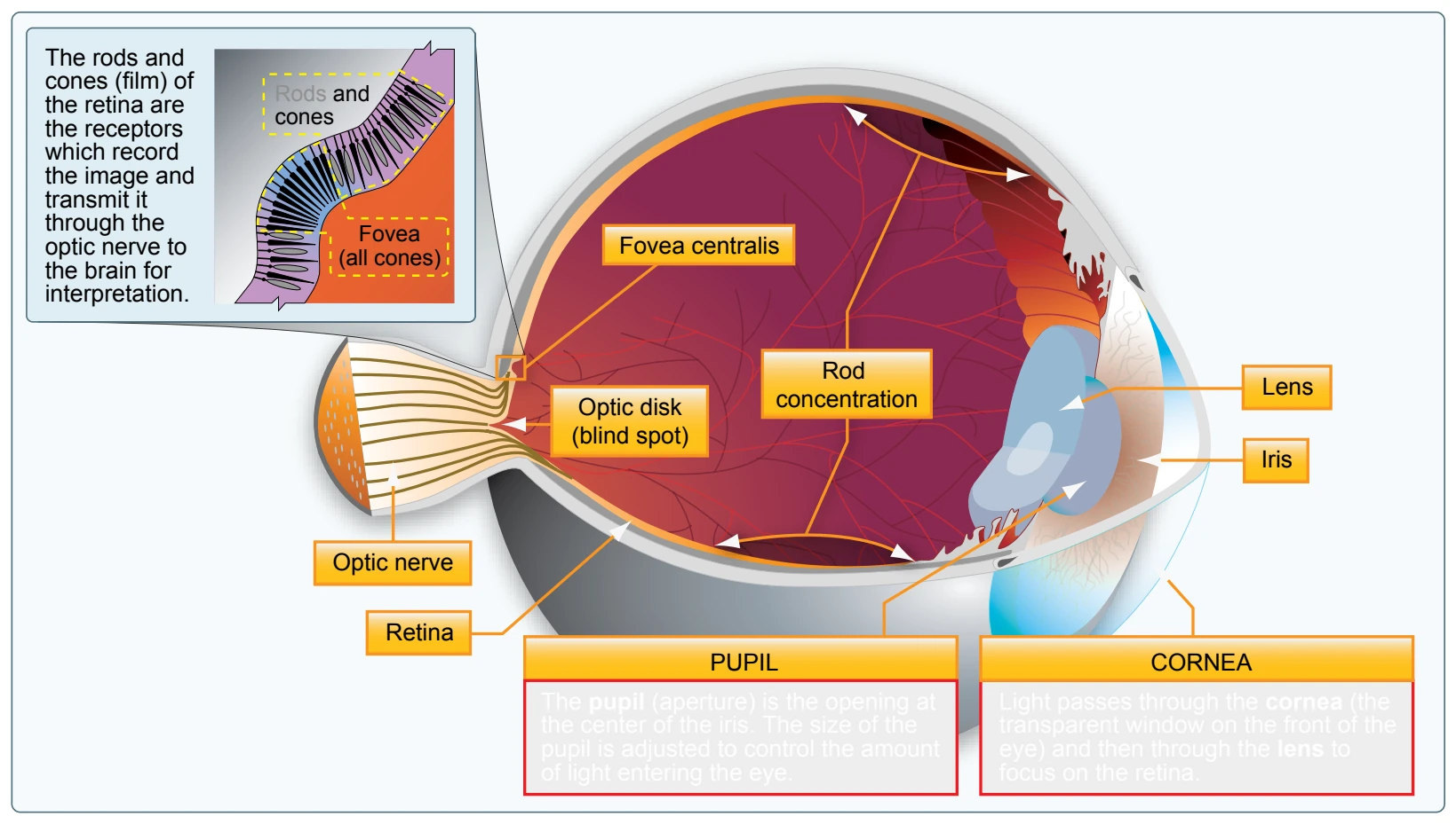
light energy into electrical impulses that travel through nerves to the brain. The brain interprets the electrical signals to form images. There are two kinds of light-sensitive cells in the eyes: rods and cones. [Figure 17-12]
The cones are responsible for all color vision, from appreciating a glorious sunset to discerning the subtle shades in a fine painting. Cones are present throughout the retina, but are concentrated toward the center of the field of vision at the back of the retina. There is a small pit called the fovea where almost all the light sensing cells are cones. This is the area where most “looking” occurs (the center of the visual field where detail, color sensitivity, and resolution are highest).
While the cones and their associated nerves are well suited to detecting fine detail and color in high light levels, the rods are better able to detect movement and provide vision in dim light. The rods are unable to discern color but are very sensitive at low-light levels. The trouble with rods is that a large amount of light overwhelms them, and they take longer to “reset” and adapt to the dark again. There are so many cones in the fovea that are at the very center of the visual field but virtually has no rods at all. So in low light, the middle of the visual field is not very sensitive, but farther from the fovea, the rods are more numerous and provide the major portion of night vision.
Vision Types
There are three types of vision: photopic, mesopic, and scotopic. Each type functions under different sensory stimuli or ambient light conditions. [Figure 17-13]
Photopic Vision
Photopic vision provides the capability for seeing color and resolving fine detail (20/20 or better), but it functions only in good illumination. Photopic vision is experienced during daylight or when a high level of artificial illumination exists.

The cones concentrated in the fovea centralis of the eye are primarily responsible for vision in bright light. [Figure 17-12] Because of the high light level, rhodopsin, which is a biological pigment of the retina that is responsible for both the formation of the photoreceptor cells and the first events in the perception of light, is bleached out causing the rod cells to become less effective.
Mesopic Vision
Mesopic vision is achieved by a combination of rods and cones and is experienced at dawn, dusk, and during full moonlight. Visual acuity steadily decreases as available light decreases and color perception changes because the cones become less effective. Mesopic viewing period is considered the most dangerous period for viewing. As cone sensitivity decreases, pilots should use off-center vision and proper scanning techniques to detect objects during low-light levels.
Scotopic Vision
Scotopic vision is experienced under low-light levels and the cones become ineffective, resulting in poor resolution of detail. Visual acuity decreases to 20/200 or less and enables a person to see only objects the size of or larger than the big “E” on visual acuity testing charts from 20 feet away. In other words, a person must stand at 20 feet to see what can normally be seen at 200 feet under daylight conditions. When using scotopic vision, color perception is lost and a night blind spot in the central field of view appears at low light levels when the cone-cell sensitivity is lost.
Central Blind Spot
The area where the optic nerve connects to the retina in the back of each eye is known as the optic disk. There is a total absence of cones and rods in this area, and consequently, each eye is completely blind in this spot. [Figure 17-14] As a result, it is referred to as the blind spot that everyone
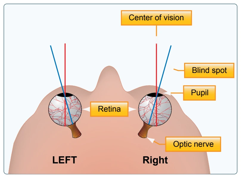
has in each eye. Under normal binocular vision conditions (both eyes are used together), this is not a problem because an object cannot be in the blind spot of both eyes at the same time. On the other hand, where the field of vision of one eye is obstructed by an object (windshield divider or another aircraft), a visual target could fall in the blind spot of the other eye and remain undetected.
- Hold this page at an arm’s length.
- Completely cover your left eye (without closing or pressing on it) using your hand or other flat object.
- With your right eye, stare directly at the airplane on the left side of the picture page. In your periphery, you will notice the black X on the right side of the picture.
- Slowly move the page closer to you while continuing to stare at the airplane.