Chapter 12
Night Operations
Visual Acuity
Normal visual acuity, or sharpness, is 20/20. A value of 20/80 indicates that an individual reads at 20 feet the letters that an individual with normal acuity (20/20) reads at 80 feet away. The human eye functions like a camera. It has an instantaneous field of view, which is oval and typically measures 120° vertically by 150° horizontally. When both eyes are used for viewing, the overall field of vision measures about 120° vertically by 200° horizontally.
The Eye
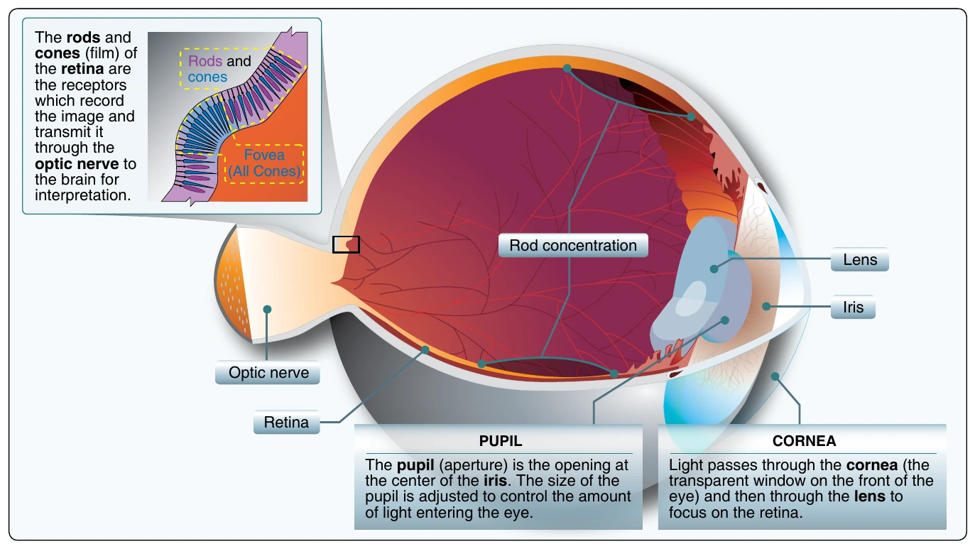
Vision is primarily the result of light striking a photosensitive layer, called the retina, at the back of the eye. The retina is composed of light-sensitive cones and rods. The cones in the eye perceive an image best when the light is bright, while the rods work best in low light. The pattern of light that strikes the cones and rods is transmitted as electrical impulses by the optic nerve to the brain where these signals are interpreted as an image.
Cones
Cones are concentrated around the center of the retina. They gradually diminish in number as the distance from the center increases. Cones allow color perception by sensing red, blue, and green light. Directly behind the lens, on the retina, is a small, notched area called the fovea. This area contains only a high concentration of cone receptors. The best vision in daylight is obtained by looking directly at the object. This focuses the image on the fovea, where detail is best seen. The cones, however, do not function well in darkness, which explains why color is not seen as vividly at night as it is during the day.
Rods
Concentrated outside the fovea area, the rods are the dim light and night receptors. The number of rods increases as the distance from the fovea increases. Rods sense images only in black and white. Because the rods are not located directly behind the pupil, they are responsible for most peripheral vision. Images that move are perceived more easily by the rod areas than by the cones in the fovea. If you have ever seen something move out of the corner of your eye, it was most likely detected by rod receptors.
In low light, the cones lose much of their function, while rods become more receptive. The eye sacrifices sharpness for sensitivity. The ability to see an object directly in front of you is reduced, and much depth perception is lost, as well as judgment of size. The concentration of cones in the fovea can make a night blindspot at the center of vision. How well a person sees at night is determined by the rods in the eyes, as well as by the amount of light allowed into the eyes. At night, the wider the pupil is open at night, the better night vision becomes.
Night Vision
Diet and general physical health have an impact on how well a person can see in the dark. Deficiencies in vitamins A and C have been shown to reduce night acuity. Other factors, such as carbon monoxide poisoning, smoking, alcohol, and certain drugs can greatly decrease night vision. Lack of oxygen can also decrease night vision as the eye requires more oxygen per unit weight than any other part of the body.
Night Scanning
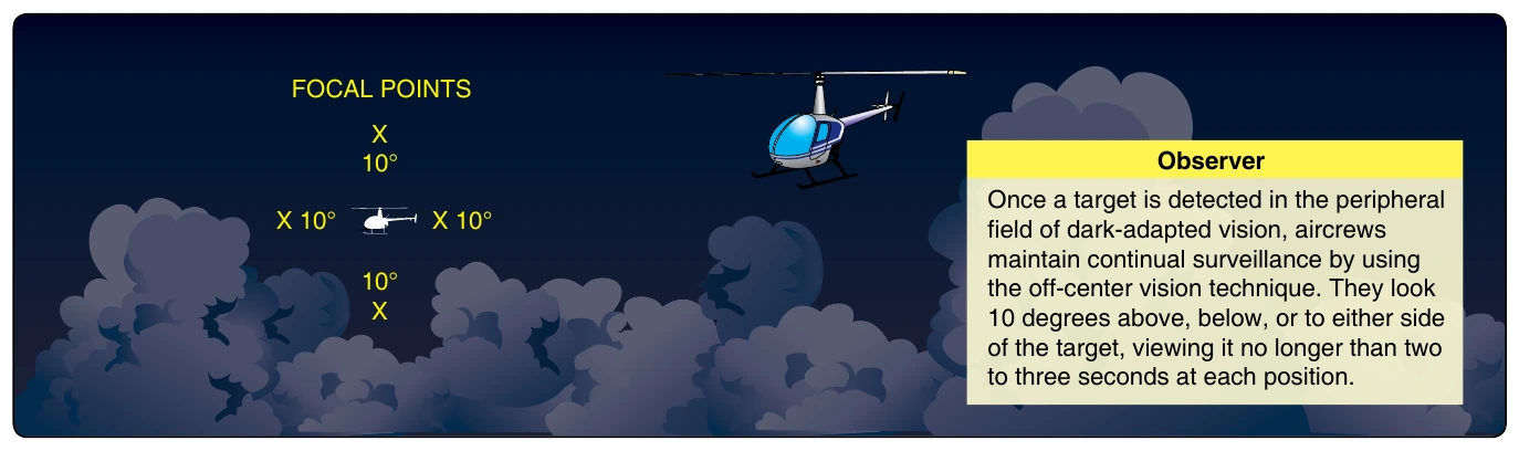
Good night visual acuity is needed for collision avoidance. Night scanning, like day scanning, uses a series of short, regularly spaced eye movements in 10° sectors. Unlike day scanning, however, off-center viewing is used to focus objects on the rods rather than the fovea blindspot. [Figure 12-4] When looking at an object, avoid staring at it too long. If staring at an object without moving the eyes, the retina becomes accustomed to the light intensity and the image begins to fade. To keep it clearly visible, new areas in the retina must be exposed to the image. Small, circular eye movements help eliminate the fading. Also, move the eyes more slowly from sector to sector than during the day to prevent blurring.
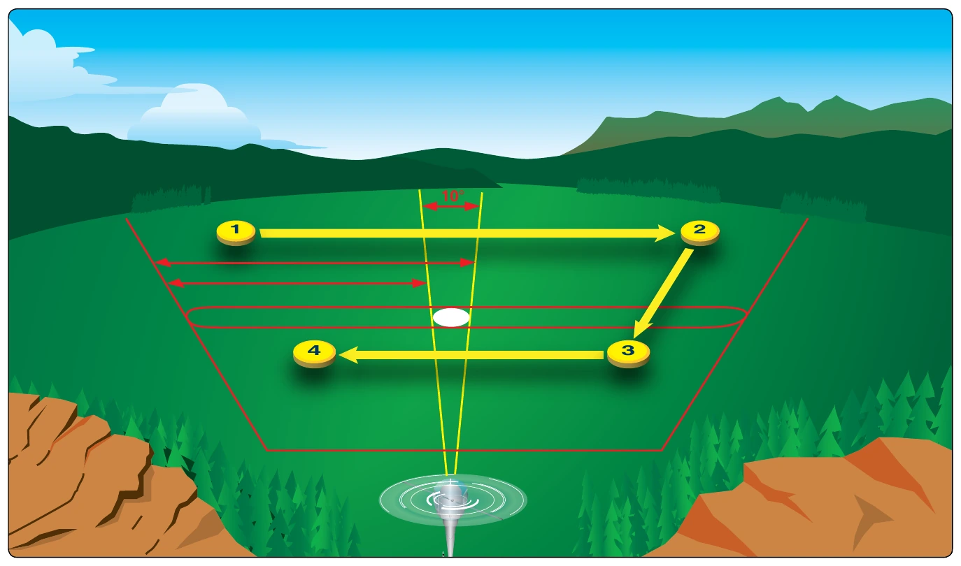
During daylight, objects can be perceived at a great distance with good detail. At night, range is limited, and detail is poor. Objects along the flight path can be more readily identified at night, by using the proper techniques to scan the terrain. To scan effectively, pilots look from side to side. They should begin scanning at the greatest distance at which an object can be perceived high on the horizon, thence moving inward toward the position of the aircraft. Figure 12-5 shows this scanning pattern. Because the light-sensitive elements of the retina are unable to perceive images that are in motion, a stop-turn-stop-turn motion should be used. For each stop, an area about 30 degrees wide should be scanned. This viewing angle includes an area about 250 meters wide at a distance of 500 meters. The duration of each stop is based on the degree of detail that is required, but no stop should last more than two or three seconds. When moving from one viewing point to the next, pilots should overlap the previous field of view by 10 degrees. This scanning technique allows greater clarity in observing the periphery. Other scanning techniques, as illustrated in Figure 12-6, may be developed to fit the situation.
Obstruction Detection
Obstructions having poor reflective surfaces, such as wires and small tree limbs, are difficult to detect. The best way to locate wires is by looking for the support structures. However, pilots should review the most current hazard maps with known wire locations before night flights.